The Flexors And Extensors Of The Wrist
Themuscleswhich flex and extend thefingersof course also move thehandas a whole, but in addition to thesemusclesthere are five others, - twoflexor musclesand threeextensor muscles, - which are inserted into thebonesof the metacarpus and not into thephalanges. When these muscles contract they tend to move the whole hand and not the fingers alone. They are the flexor carpi radialis, flexor carpi ulnaris, extensor carpi radialis longior, extensor carpi radialis brevior, and extensor carpi ulnaris. The palmaris longus has already been described as aflexorof the fingers.
Flexors Of The Wrist
Flexor Carpi Radialis
The two flexors of the wrist, the flexor carpi radialis and the flexor carpi ulnaris, are bothsuperficialmuscles lying directly beneath theskin. The flexor carpi radialis arises from the medial (internal) condyle of thehumerusandintermuscular septaand lies between thepronatorradii teresexternallyand the palmaris longus internally. It runs obliquely across theforearm, striking the wrist at about the junction of the middle and outer thirds. It lies next to and to the outer side of the palmaris longustendonand to the ulnar side of theradial arteryand inserts into the front of the base of the secondmetacarpal bone(Fig. 324).
Flexor Carpi Ulnaris
The flexor carpi ulnaris arises by two heads, one from the common tendon of the medial (internal) condyle and the other from the olecranon process and upper two-thirds of theulna. The two heads are separated by theulnar nerve, which passes down in the groove between the medial condyle and olecranon process. The muscle passes straight down theanteriorand inner surface of the ulna to insert first into the pisiform bone and unciform process and then to continue over to the base of the fifth metacarpal bone. The pisiform bone is a sesamoid bone in the tendon of the flexor carpi ulnaris muscle.
Both the flexor carpi radialis and the flexor carpi ulnaris flex the hand at the wrist. When the ulnaris alone acts it tends to tilt the hand inward; when the radialis acts alone it tends to incline the hand outward. Being superficial, these muscles are both important landmarks and guides to thearteries.

Fig. 324. - The flexor muscles of the wrist.

Fig. 325. - The extensor muscles of the wrist.
Extensors Of The Wrist
Extensor Carpi Radialis Longior
The extensor carpi radialis longior arises from the lower third of theexternalsupracondylarridge and the lateral (external) condyle and inserts into thebackof the base of the second metacarpal bone. When it contracts it tends to tilt the hand toward the radial side as well as to extend it, and, being attached to the humerus above thelineof theelbow-joint, it also aids in flexing the elbow.
Extensor Carpi Radialis Brevior
The extensor carpi radialis brevior arises from the common tendon of the lateral condyle andfascia, and, running down parallel to the longior muscle, inserts into the base of the third metacarpal bone. It is covered by the extensor carpi radialis longior muscle and lies on the supinator (brevis). It acts as a pureextensorof the wrist (Fig. 325).
Extensor Carpi Ulnaris
The extensor carpi ulnaris arises by two heads, one from the lateral (external) condyle and the other from theposterior surfaceof the ulna through the fascia common to it, to the flexor carpi ulnaris, and to the flexor profundus digitorum. It inserts into the base of the fifth metacarpal bone. It extends the wrist and tilts the hand toward the ulnar side.
http://chestofbooks.com/health/anatomy/Human-Body-Construction/2-The-Flexors-And-Extensors-Of-The-Wrist.html
<><<
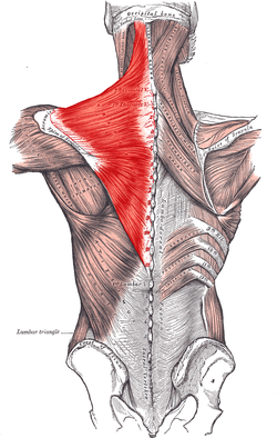
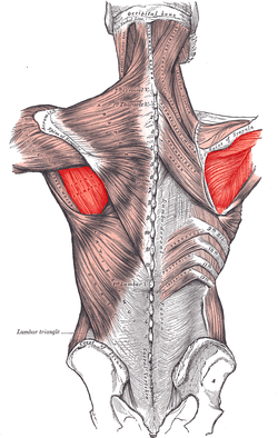
In human anatomy, the trapezius (/trəˈpiːzi.əs/) is one of two large superficial muscles that extend longitudinally from the occipital bone to the lower thoracic vertebrae and laterally to the spine of thescapula (shoulder blade). Its functions are to move the scapulae and support the arm.
The trapezius has three functional regions: the superior region (descending part), which supports the weight of the arm; the intermediate region (transverse part), which retracts the scapulae; and the inferior region (ascending part), which medially rotates and depresses the scapulae.

https://en.wikipedia.org/wiki/Trapezius_muscle
<><
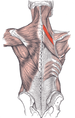
The rhomboid major is a skeletal muscle on the back that connects the scapula with the vertebrae of thespinal column. In human anatomy, it acts together with the rhomboid minor to keep the scapula pressed against thoracic wall and to retract the scapula toward the vertebral column.[1]

In human anatomy, the rhomboid minor is a small skeletal muscle on the back that connects the scapulawith the vertebrae of the spinal column.
Located inferior to levator scapulae and superior to rhomboid major, it acts together with the latter to keep the scapula pressed against the thoracic wall. It lies deep to trapezius but superficial to the long spinal muscles.[1]
https://en.wikipedia.org/wiki/Rhomboid_minor_muscle
<><>
Anatomy and supply. The levator scapulae is a longmuscle of the shoulder girdle. It originates at the transverse processes of the atlas and axis as well as the posterior tubercles of the 3rd-4th cervical vertebrae. It inserts at the superior angle and medial border of thescapula.

https://www.google.com.au/search?sclient=psy-ab&biw=1197&bih=831&q=trapezius+muscles&oq=trape+muscles&gs_l=serp.1.0.0i7i30k1l4.2676.7432.0.10486.5.5.0.0.0.0.299.1303.2-5.5.0....0...1c.1.64.psy-ab..1.4.977...0i67k1.9CwJZwHp_zM&pbx=1&dpr=1&cad=cbv&bvch=u&sei=ZirNV7CKC8a_0gTFipXIBw#q=levator+scapulae+muscles
<><
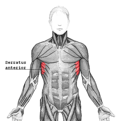
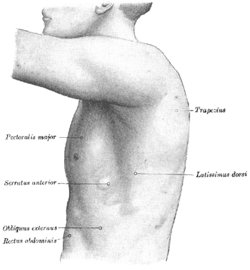
The serratus anterior (/ˌsᵻˈreɪtəs ænˈtɪəri.ər/) (Latin: serrare = to saw, referring to the shape, anterior = on the front side of the body) is a muscle that originates on the surface of the 1st to 8th ribs at the side of the chest and inserts along the entire anterior length of the medial border of the scapula.
The left side of the thorax.
https://en.wikipedia.org/wiki/Serratus_anterior_muscle
<><
The latissimus dorsi (/ˌləˈtɪsᵻməs ˈdɒrsaɪ/) (plural: latissimi dorsi), meaning 'broadest [muscle] of the back' (Latin latus meaning 'broad', latissimus meaning 'broadest' and dorsum meaning the back), is the larger, flat, dorso-lateral muscle on the trunk, posterior to the arm, and partly covered by the trapezius on its median dorsal region. Latissimi dorsi are commonly known as "lats", especially among bodybuilders.
The latissimus dorsi is responsible for extension, adduction, transverse extension also known as horizontal abduction, flexion from an extended position, and (medial) internal rotation of the shoulder joint. It also has a synergistic role in extension and lateral flexion of the lumbar spine.
Due to bypassing the scapulothoracic joints and attaching directly to the spine, the actions the latissimi dorsi have on moving the arms can also influence the movement of the scapulae, such as their downward rotation during a pull up.
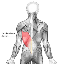

https://en.wikipedia.org/wiki/Latissimus_dorsi_muscle
<><<
The pectoralis major (/ˌpɛktəˈreɪlᵻs ˈmeɪdʒər/) (from Latin: pectus, breast) is a thick, fan-shaped muscle, situated at the chest (anterior) of the human body. It makes up the bulk of the chest muscles in the male and lies under the breast in the female. Underneath the pectoralis major is the pectoralis minor, a thin, triangular muscle. In sports as well as bodybuilding, the pectoral muscles may colloquially be referred to as "pecs", "pectoral muscle" or "chest muscle".

Superficial muscles of the chest and front of the arm.
<><<
n human anatomy, the deltoid muscle is the muscle forming the rounded contour of the shoulder. Anatomically, it appears to be made up of three distinct sets of fibers though electromyography suggests that it consists of at least seven groups that can be independently coordinated by the nervous system.[1]
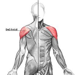

https://en.wikipedia.org/wiki/Deltoid_muscle
,.>
The supraspinatus (plural supraspinati, from Latin supraspinatus) is a relatively small muscle of the upper back that runs from the supraspinatous fossa superior of the scapula (shoulder blade) to thegreater tubercle of the humerus. It is one of the four rotator cuff muscles and also abducts the arm at the shoulder. The spine of the scapula separates the supraspinatus muscle from the infraspinatus muscle, which originates below the spine.

https://en.wikipedia.org/wiki/Supraspinatus_muscle
<><
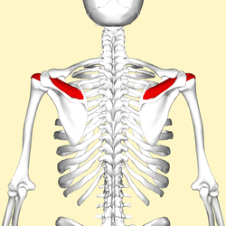
The teres major muscle (Latin teres meaning 'rounded') is a muscle of the upper limb and one of sevenscapulohumeral muscles. It is a thick but somewhat flattened muscle, innervated by the lower subscapular nerve (C5 and C6).

<><
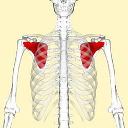
The teres minor (Latin teres meaning 'rounded') is a narrow, elongated muscle of the rotator cuff.

<><
<><
The teres minor (Latin teres meaning 'rounded') is a narrow, elongated muscle of the rotator cuff.
<><
The subscapularis is a large triangular muscle which fills the subscapular fossa and inserts into thelesser tubercle of the humerus and the front of the capsule of the shoulder-joint.
<><
In human anatomy, the infraspinatus muscle is a thick triangular muscle, which occupies the chief part of the infraspinatous fossa.[1] As one of the four muscles of the rotator cuff, the main function of the infraspinatus is to externally rotate the humerus and stabilize the shoulder joint.
<><<
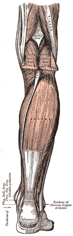
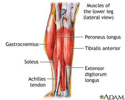
In humans, the gastrocnemius muscle (/ˌɡæstrɒkˈniːmiəs/ or /ˌɡæstrəkˈniːmiəs/; plural gastrocnemii;Latin, from Greek γαστήρ "stomach" and κνήμη (knēmē) "leg"; meaning "stomach of leg" (referring to the bulging shape of the calf) is a very powerful superficial bipennate muscle that is in the back part of the lower leg. It runs from its two heads just above the knee to the heel, a two joint muscle.

<><<>
In humans and some other mammals, the soleus is a powerful muscle in the back part of the lower leg(the calf). It runs from just below the knee to the heel, and is involved in standing and walking. It is closely connected to the gastrocnemius muscle and some anatomists consider them to be a single muscle, thetriceps surae. Its name is derived from the Latin word "solea", meaning "sandal".
<><>
 <><
<><


In human anatomy, the infraspinatus muscle is a thick triangular muscle, which occupies the chief part of the infraspinatous fossa.[1] As one of the four muscles of the rotator cuff, the main function of the infraspinatus is to externally rotate the humerus and stabilize the shoulder joint.
<><<
In humans, the gastrocnemius muscle (/ˌɡæstrɒkˈniːmiəs/ or /ˌɡæstrəkˈniːmiəs/; plural gastrocnemii;Latin, from Greek γαστήρ "stomach" and κνήμη (knēmē) "leg"; meaning "stomach of leg" (referring to the bulging shape of the calf) is a very powerful superficial bipennate muscle that is in the back part of the lower leg. It runs from its two heads just above the knee to the heel, a two joint muscle.
<><<>
In humans and some other mammals, the soleus is a powerful muscle in the back part of the lower leg(the calf). It runs from just below the knee to the heel, and is involved in standing and walking. It is closely connected to the gastrocnemius muscle and some anatomists consider them to be a single muscle, thetriceps surae. Its name is derived from the Latin word "solea", meaning "sandal".
<><>
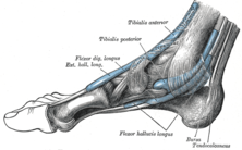
In human anatomy, the tibialis anterior (/ˌtɪbiˈeɪlᵻs/ or /ˌtɪbiˈælᵻs/) is a muscle that originates in the upper two-thirds of the lateral (outside) surface of the tibia and inserts into the medial cuneiform and firstmetatarsal bones of the foot. It acts to dorsiflex and invert the foot. This muscle is mostly located near the shin.
It is situated on the lateral side of the tibia; it is thick and fleshy above, tendinous below. The tibialis anterior overlaps the anterior tibial vessels and deep peroneal nerve in the upper part of the leg.
The tibialis posterior is the most central of all the leg muscles, and is located in the deep posterior compartment of the leg.
It is the key stabilizing muscle of the lower leg.
Blood is supplied to the muscle by the posterior tibial artery, and innervation is via the tibial nerve.
<><>
In human anatomy, the peroneus longus (also known as fibularis longus) is a superficial muscle in the lateral compartment of the leg, and acts to evert and plantar flex the ankle.
<><<
<>>
<><
<><<
<><
<><<
<><
<><
tensor fasciae latae muscles
<>
adductor muscles
<>>
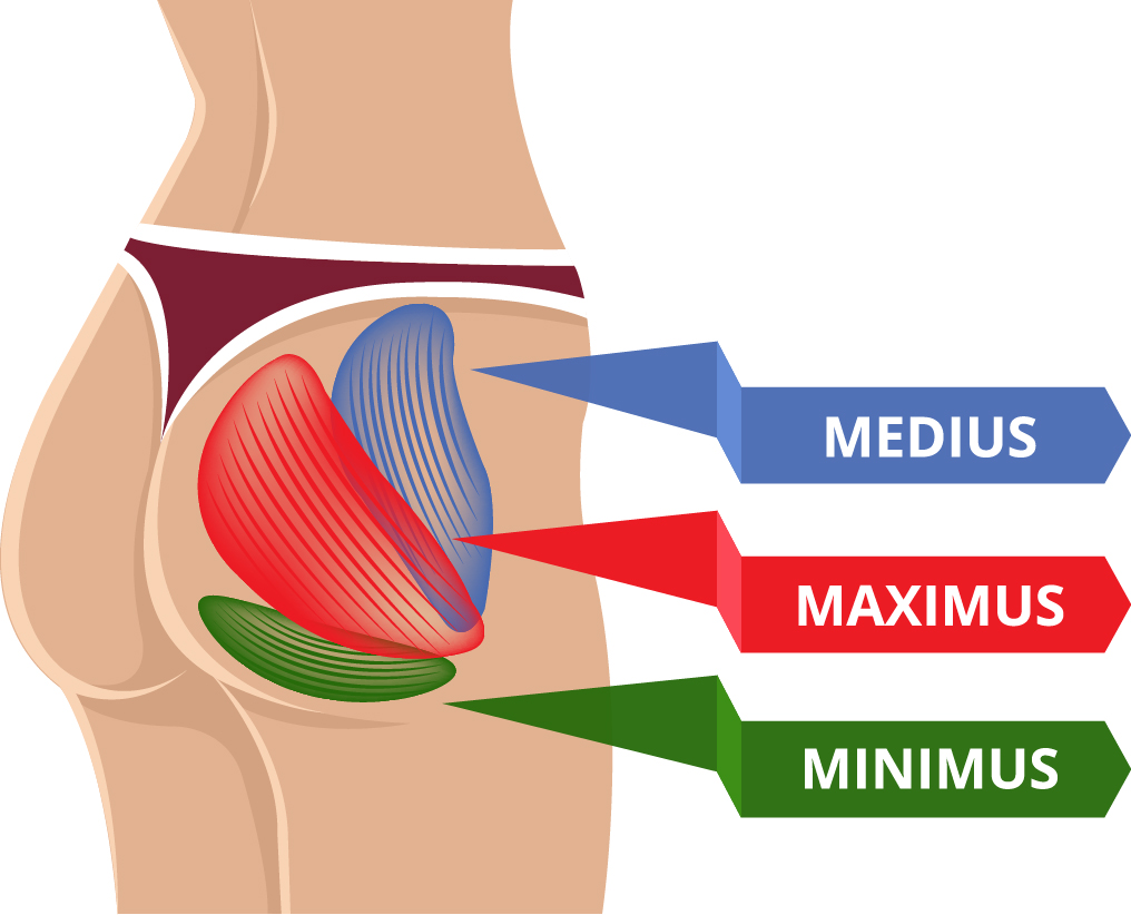
gluteus muscles
<><
Piriformis muscles
<><
psoas muscles
<>
Iliacus muscles.
<><
abdominal muscles
,.<><.,
Flexors-And-Extensors-Of-The-Wrist.html#ixzz4JMpz3Us0
No comments:
Post a Comment